- Varicose veins treatment in Kyiv
- Varicose veins treatment in Zaporizhzhia
- Edema and lymphostasis treatment
- Myths about varicose veins
- Massage and varicose veins
- Treatment of varicose veins with hydrogen peroxide
- Veins ache and pull blood vessels
- Can I treat varicose veins with pills?
- Treatment for varicose veins with leeches and bees. Benefit or harm?
- Is it possible to get rid of varicose veins with the help of gels and ointments?
- After 70 years, is it too late to treat varicose veins?
- Do mud baths help with varicose veins?
- Is it possible to get rid of varicose veins with compression knitwear?
- If you make an "open" operation - varicose veins will never appear again. Is it so?
- Are ugly scars and scars left after the operation to remove varicose veins?
- Is varicose veins treatment a long and painful process?
- Is it possible to get rid of varicose veins completely and forever?
- Varicose veins cannot be treated in the summer. Is it so?
- Do you need to go to a surgical hospital for vein surgery or not?
- After removal of varicose veins, the load on healthy veins increases. Is this so?
- Is it possible to play sports after surgery to remove varicose veins?
- Only the elderly suffer from varicose veins?
- Wearing compression hosiery causes muscle atrophy or not?
- Remove veins or not?
- How is the outflow of blood after vein removal?
- Thrombophlebitis treatment
- Nonresident patients
- All about varicose veins
- The risk of complications of varicose veins | Trophic ulcers and no panic
- Causes and symptoms of varicose veins
- Diagnosis of varicose veins
- Stage of development of varicose veins
- Gymnastics and prevention of varicose veins
- Diet for varicose veins
- Why is varicose veins dangerous?
- Treatment of varicose veins of the lower extremities
- Varicose veins and pregnancy
- Varicose disease - what will happen if not treated?
- Varicose symptoms
- Complications of endovenous treatments
- Post-thrombotic disease
- What is phlebology?
- Complications of varicose veins
- All about sclerotherapy
- Vascular diseases
- Treatment results in AngioLife
- Questionnaires and tests for patients and doctors
- For doctots
- Public contract
Order a call
Diagnosis, clinical and treatment of varicose veins
08 feb 2025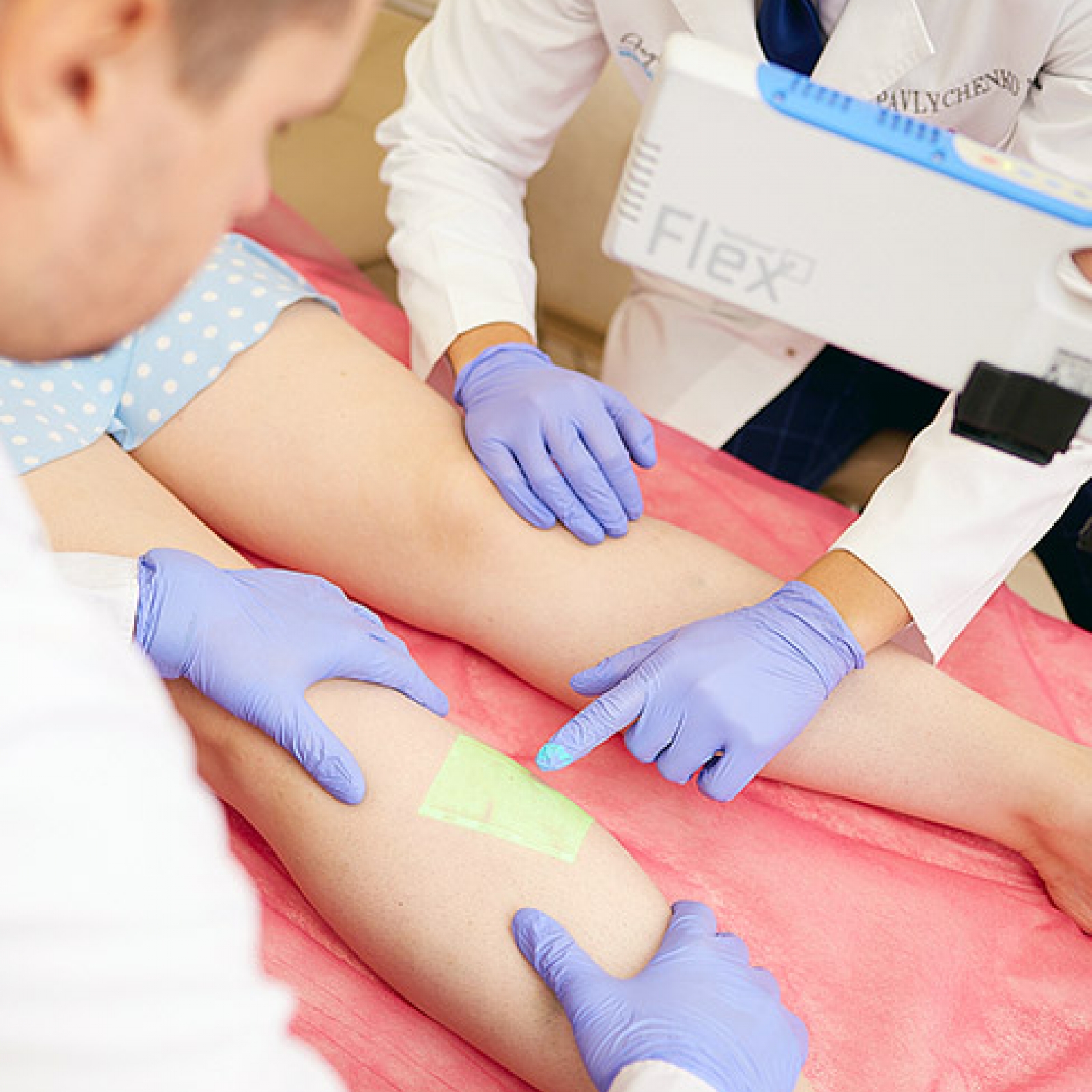
Varicose veins are dilated, visible superficial veins resulting from venous insufficiency, which mainly affect the lower extremities of a person. The movement of blood through the veins occurs from the bottom up and is based on the work of one-way valves and the calf muscle pump, the incorrect operation of which leads to venous reflux and venous hypertension.
Pathophysiologically, venous reflux disrupts normal blood flow, increasing the stretching of the vein walls. Valvular insufficiency of the perforating veins and other types of chronic venous insufficiency (CVI) are often a precursor to varicose veins. CVI is manifested by increased venous pressure, inflammation and impaired microcirculation, which can potentially progress to venous ulcers. Further expansion of the veins usually ends in varicose veins (venous hypertension). Today, superficial venous insufficiency occurs in 50–60% of adults in Western countries, which has a significant socio-economic impact, including due to the costs associated with treatment.
Venous disease in general is characterized by the occurrence of serious complications, including, in particular: inflammation of the veins (phlebitis), varicose vein thrombosis, thromboembolism (PE), chronic thromboembolic pulmonary hypertension and a high risk of mortality.
Etiology of varicose veins
What are the causes of varicose veins? The following risk factors contribute to its development:
Genetic predisposition (a family history of varicose veins significantly increases the susceptibility to the disease);
Older age, which leads to degenerative changes in the walls and valves of the veins;
Women are more susceptible to the disease, especially during menopause;
Increased blood volume, hormonal changes and pressure in the uterus during pregnancy can also provoke varicose veins;
Occupations or lifestyle factors that require prolonged periods of immobility: sitting or standing, which impede venous outflow, and a generally sedentary lifestyle;
Excess body weight puts additional pressure on the veins;
Having nonthrombotic iliac vein obstruction (May-Turner syndrome);
Trauma and surgery can disrupt venous flow and valve function;
A history of deep vein thrombosis (DVT) changes the structure of the veins, increasing the risk of superficial vein dilation;
Smoking;
Use of oral contraceptives.
Clinical presentation of varicose veins
Patients with varicose veins may have a number of symptoms:
Prominent, bluish, tortuous, visible veins in the legs;
Pain or a feeling of heaviness in the legs, often worse after prolonged standing or sitting;
Swelling, especially around the ankles due to fluid retention;
Hyperpigmentation or pale atrophy of the skin due to hemosiderin deposition when red blood cells are extravasated into the surrounding tissues;
Venous eczema, lipodermatosclerosis due to fibrosis of the subcutaneous fat;
Chronic venous insufficiency can lead to venous ulcers, usually around the medial malleolus.
The severity of symptoms does not always correspond to the apparent degree of varicose veins, which requires a careful diagnostic examination.
How to diagnose varicose veins
Effective diagnosis of varicose veins involves a combination of patient history, physical examination, and modern diagnostic methods:
Study of the medical history after obtaining information about the onset of symptoms, duration, aggravating factors, family history, occupational characteristics, and previous interventions.
Physical examination, which includes examining the patient in a standing position for visible varicose veins, skin discoloration, ulcers, and palpation for tenderness, induration, or arteriovenous malformation.
Functional tests such as the Trendelenburg test to assess the efficiency of the saphenofemoral junction and superficial veins and the Perthes test to assess the functionality of the deep venous system.
Duplex/Triplex Ultrasound (Vascular Ultrasound). Visualization of the anatomy of the veins and examination of blood flow and valve function are performed. This allows for the detection of reflux, obstruction and thrombosis of the deep veins.
Photoplethysmography (PPG) to measure venous filling time (to assess venous function).
Magnetic resonance venography (MRV) to obtain a detailed image in cases where evaluation of the venous structures of the pelvis is required.
Invasive venography, which involves the injection of contrast to visualize the venous anatomy. It is prescribed in complex cases or for preoperative planning. Invasive diagnostic and treatment procedures are not performed in our clinic.
In addition, differential diagnosis allows for the diagnostic separation of varicose veins from other possible diseases, such as deep vein thrombosis, peripheral artery disease, lymphedema, and others.
Why should varicose veins be treated
If varicose veins are not treated, they can lead to:
Chronic venous insufficiency (persistent venous hypertension that causes skin changes). The prevalence of chronic venous diseases in the world reaches 83.6%, and in Ukraine up to 20% of the population suffer from varicose veins;
Venous ulcers: painful, non-healing wounds, usually above the medial malleolus;
Superficial thrombophlebitis (inflammation and thrombosis of the superficial veins, manifested by erythema and tenderness along the vein);
Bleeding due to trauma to superficial varicose veins.
What offers effective treatment of varicose veins in our clinic
The basis of all treatment of varicose veins is the elimination of pathological venous reflux in the main subcutaneous veins.
We focus only on non-surgical treatment, we offer our patients, first of all, innovative conservative measures: restoration of incompetent valves, phlebological compression stockings, special functional exercises, as well as pharmacological treatment, which includes taking venoactive drugs to improve vein tone. Direct non-invasive treatment includes endovenous procedures: sclerotherapy, bioglue, radiofrequency vein ablation and laser treatment and cryolaser-cryosclerotherapy.
References:
1. Singer PN. Galen. In: Edward NZ (ed). The Stanford Encyclopedia of Philosophy, Stanford, CA: Metaphysics Research Lab Philosophy Department, Stanford Univer sity Stanford. 2021 Edition
2. Tepelenis K, Papathanakos G, Kitsouli A, Barbouti A, Varvarousis DN, Kefalas A, et al. Anatomical variations of the great saphenous vein at the saphenofemoral junction. A cadaveric study and narrative review of the literature. Vascular. 2023
3. Langford JT, Dardik A. Vessel wall biology. In: Sidawy AN, Perler BA, editors. Rutherford's Vascular Surgery and Endovascular Therapy. Philadelphia: Elsevier. 2023. 29–40
Latest materials
- ANGIOLIFE at the UVS Congress - Sukharev Readings 2025
- Thromboangiitis obliterans, Buerger's disease and Endarteritis
- Тромбофлебіт та тромбоз поверхневих вен
- ANGIOLIFE on aspects of phlebology 2024
- Is it possible to treat deep vein thrombosis without anticoagulant therapy?
- UVS 2024 CONGRESS "SUKHAREV READINGS"
- Varicose veins
- Nutcracker Syndrome
Get a consultation
about services by phone
about services by phone
Has any questions?
Fill out the form and we will call you back!